The confocal microscope is really good.
recently discovered a previously unnoticed award for scientific photography, sponsored by Cancer Research UK (ICR), called the ICR Science and Medical Imaging Competition (ICR Science and Medical Imaging competition). When I happened to see it, I would like to introduce some award-winning works of this competition.
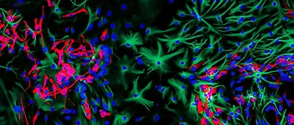
the winning work selected by the jury this year came from Sumana Shrestha, a doctoral student. This microphotograph shows the differentiation of neural stem cells from mice. These cells are used in the laboratory to study glioblastoma, a highly aggressive brain tumor.
this image was taken with a confocal microscope. Different cells were distinguished by immunostaining. Astrocytes were marked green and neurons were marked red. The cells used in the study were genetically edited and then treated with viruses to induce tumors as a research model.
while followers of ICR social media accounts like the following one, also from Sumana Shrestha. This electron microscope image shows some nanoparticles with a diameter about the size of a human hair. Nanoparticles can be used to deliver drugs, and the surface microstructure of nanoparticles can also affect the response of cells. Some netizens think that these nanoparticles look a bit like pollen.
the following two award-winning works
metastatic mouse melanoma cells spread on the collagen matrix and were imaged by total internal reflection fluorescence (TIRF) microscope. Authors: Dr Chris Bakal and Oliver Inge
Buy our special occasion dress shops in ontario and enjoy the combination of their high quality and superior material. Shop now and enjoy the pleasant shopping experience.
cancer cells (pink) invaded a layer of vascular cells and were photographed by confocal microscopy. Author: Dr. Maxine Lam
generally speaking, sensory cells look good as long as they are photographed by fluorescence-labeled confocal microscopy (
Source: https://www.icr.ac.uk/news-features/latest-features/the-art-of-science-cutting-edge-science-and-captivating-images